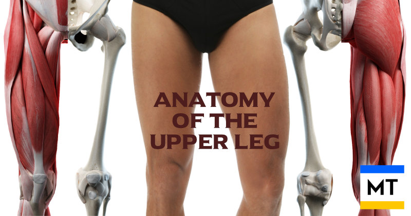


Understanding the anatomy and dynamics of the upper leg is vital for athletes, physical therapists, and students of musculoskeletal anatomy. Let's explore the function, origins, and insertion points of the bones and muscles involved in the upper leg.
Enjoy the article. Feel free to contact me if you have any questions, or drop into my Rockhampton studio for a chat. My details are on the bookings page.
Have a brilliant day,
Andrew Thompson
![]()
![]()
![]()
![]()

Bones of the upper leg
The upper leg spans from the hip joint to the knee joint, and its structure is centred around one major bone - the femur.
The femur is the longest and strongest bone in the body. It supports body weight, serves as an attachment for muscles, and connects with adjacent bones: the pelvis at the hip joint above and the tibia and patella at the knee joint below.
The head of the femur (or femoral head) articulates with the acetabulum of the pelvis, forming the hip joint, while its distal end forms the upper part of the knee joint.
Fun fact: The femur is capable of withstanding forces of 1800 - 2500 pounds per square inch before fracturing 😲

Muscles of the upper leg
The muscles of the upper leg are divided into three compartments: anterior (front), medial (inner thigh), and posterior (back). Each compartment contains muscles that contribute to movement and stability.
Quadriceps (Anterior Compartment)
The quads comprises four muscles located anterior to the femur. Their roles include knee extension and hip flexion.
Rectus Femoris
Originates at the anterior inferior iliac spine and inserts at the tibial tuberosity via the patellar tendon. It extends the knee and flexes the hip.
Vastus Lateralis
Originates at the greater trochanter and linea aspera of the femur, inserts at the tibial tuberosity. Responsible for knee extension.
Vastus Medialis
Originates at the medial lip of the linea aspera, inserts at the tibial tuberosity. Extends the knee and stabilises the patella.
Vastus Intermedius
Originates at the anterior and lateral surfaces of the femur, inserts at the tibial tuberosity. Extends the knee.
Sartorius (Medial Compartment)
The longest muscle in the body, it originates at the anterior superior iliac spine and inserts at the medial surface of the tibia. It flexes, abducts, and laterally rotates the hip, and flexes the knee.
Fun Fact: Named from the Latin for "tailor", referencing the crossed-leg position tailors would sit in, this muscle helps achieve that position.

Adductors (Medial Compartment)
Adductors are a group of muscles that adduct the hips, and also medially rotate the thighs (rotate inwards), working as antagonists to the glutes.
Adductor Longus, Brevis, and Magnus
These muscles originate from the pubis and insert along the femur's linea aspera and adductor tubercle. They adduct and medially rotate the thigh.
Pectineus
Pectineus originates at the superior pubic ramus, inserts at the pectineal line of the femur. Adducts and flexes the hip.
Gracilis
Gracilis originates at the inferior pubic ramus and inserts at the medial tibia. It adducts the thigh and flexes the knee.
Hamstrings (Posterior Compartment)
The muscles collectively called the hamstrings, are located behind the femur. They serve to flex the knee and extend the hip.
Biceps Femoris
"Biceps" means 'two heads'. The long head of Biceps Femoris originates at the ischial tuberosity, while the short head originates at the linea aspera of the femur. The muscle inserts at the head of the fibula. Biceps Femoris flexes the knee and extends the hip.
Semitendinosus
Semitendinosus originates at the ischial tuberosity and inserts at the medial tibia. It extends the hip and flexes the knee.
Semimembranosus
Semimembranosus also originates at the ischial tuberosity, but inserts on the medial tibial condyle. Semimembranosus extends the hip and flexes the knee.
Key nerves and blood vessels of the upper leg
- Femoral nerve (anterior compartment) innervates the quadriceps and supplies skin sensation.
- Obturator nerve (medial compartment) innervates the adductors.
- Sciatic nerve (posterior compartment) innervates the hamstrings and lower leg.
- Femoral artery is a continuation of the external iliac artery, and provides arterial blood to the upper leg.