
Core muscles wrap around our torsos like a coccoon, working together to keep our bodies upright, protect our internal organs, and transmit force between the upper and lower body.
Understanding how the core muscles work with and against each other is crucial for athletes, trainers, remedial therapists, physiotherapists, and students of functional anatomy.
Enjoy the article. Feel free to contact me if you have any questions, or drop into my Rockhampton studio for a chat. My details are on the bookings page.
Have a brilliant day,
Andrew Thompson
![]()
![]()
![]()
![]()

Muscle Core divisions
Based on their location, muscles of the human core are generally grouped into areas as below. Based on function though, core muscles can belong to multiple groups.
Anterior abdominals
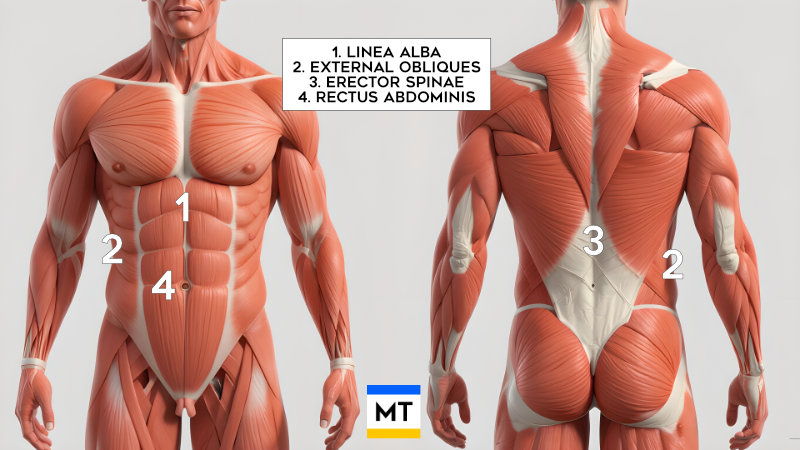
Rectus abdominis
Rectus Abdominis is a paired muscle, in that it is divided vertically by the linea alba, a band of connective tissue which sits in front of the muscle. In people with low body fat, the linea alba is visible as a vertical groove.
The "abs" flex the vertebral column as in performing a sit-up. It also compresses abdominal contents and stabilises the pelvis when walking.
- Origin: Pubic crest and pubic symphysis
- Insertion: Xiphoid process of sternum and costal cartilages of ribs 5-7
Pyramidalis
The pyramidalis tenses the linea alba. Not everyone has a pyramidalis.
- Origin: Pubic crest
- Insertion: Linea alba
Lateral abdominals
External oblique
The external obliques bilaterally flex the trunk. Unilaterally, they rotate the trunk to the opposite side, and laterally flex the trunk. They also compress the abdominal viscera.
- Origin: Outer surfaces of ribs 5-12
- Insertion: Linea alba, pubic tubercle, anterior half of iliac crest
Internal oblique
The internal obliques bilaterally flex the trunk. Unilaterally, they rotate the trunk to the same side, and laterally flex the trunk. They also compress the abdominal viscera.
- Origin: Thoracolumbar fascia, anterior two-thirds of iliac crest, lateral half of inguinal ligament
- Insertion: Inferior borders of ribs 10-12, linea alba, pubis via conjoint tendon
Transversus abdominis
The transversus abdominis is a deeper lateral abdominal muscle. It compresses and supports the abdominal viscera, and provides deep core stabilisation.
- Origin: Internal surfaces of costal cartilages 7-12, thoracolumbar fascia, iliac crest, lateral third of inguinal ligament
- Insertion: Linea alba with aponeurosis of internal oblique, pubic crest, and pecten pubis via conjoint tendon
Posterior core muscles
Erector spinae group
The erector spinae group comprises the iliocostalis, longissimus, and spinalis muscles. They extend the vertebral column, laterally flex the vertebral column, and also control posture.
- Origin: Broad tendon from posterior iliac crest, sacrum, sacroiliac ligaments, sacral and lumbar spinous processes, supraspinous ligament.
- Insertion: Iliocostalis — ribs and cervical transverse processes; Longissimus — ribs, transverse processes, mastoid process; Spinalis — spinous processes in upper thoracic and cervical regions
Multifidus
The multifidus stabilises vertebrae during local movements of the vertebral column.
- Origin: Sacrum, posterior superior iliac spine, mammillary processes of lumbar vertebrae, transverse processes of thoracic vertebrae, articular processes of C4-C7
- Insertion: Spinous processes of vertebrae 2-4 segments above
Quadratus lumborum
The quadratus lumborum laterally flexes the vertebral column, fixes rib 12 during inspiration, and assists in extension.
- Origin: Medial half of inferior border of rib 12, tips of lumbar transverse processes
- Insertion: Iliolumbar ligament and internal lip of iliac crest
Pelvic floor
Levator ani group
The levator ani group comprises the puborectalis, pubococcygeus, and iliococcygeus muscles. They support pelvic viscera, maintain continence, and assist in core stability.
- Origin: Body of pubis, tendinous arch of obturator fascia, ischial spine
- Insertion: Perineal body, coccyx, walls of prostate or vagina, rectum, and anal canal
Coccygeus (Ischiococcygeus)
the coccygeus supports the pelvic viscera, and flexes the coccyx.
- Origin: Ischial spine
- Insertion: Inferior sacrum and coccyx
Diaphragm
The primary function of the diaphragm muscle is respiration, however it also contributes to intra-abdominal pressure for core stabilisation.
- Origin: Xiphoid process, internal surfaces of lower six ribs and costal cartilages, lumbar vertebrae via crura
- Insertion: Central tendon
Summary
The core is an integrated system of deep and superficial muscles working together to stabilise, protect, and mobilise the torso. They anchor movement, aid breathing, and safeguard the spine during activity.